
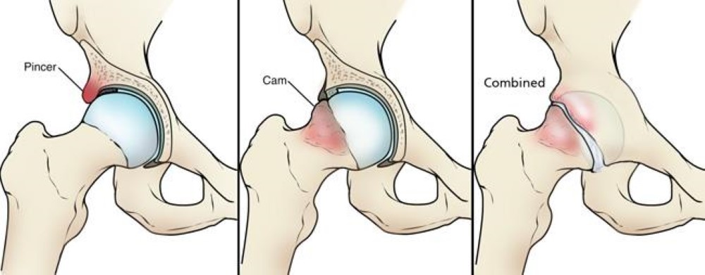
Femoroacetabular impingement, commonly referred to as FAI, is a condition characterized by abnormal contact between the femoral head and the acetabulum of the hip joint. This interaction leads to a variety of symptoms including pain, limited range of motion, and can progressively result in osteoarthritis if left untreated. As its name suggests, FAI is an impingement issue that primarily occurs due to structural abnormalities of the hip joint, which can manifest in diverse forms.
There are three primary types of femoroacetabular impingement: cam impingement, pincer impingement, and mixed impingement, each displaying distinct characteristics that can affect individuals differently. Understanding these types is crucial for accurate diagnosis and appropriate treatment.
1. Cam Impingement: In cam impingement, the femoral head is not perfectly spherical, which leads to abnormal contact with the acetabulum during leg movements. This irregularity can cause the femoral head to ‘catch’ on the edge of the acetabulum, resulting in pain and decreased mobility. Individuals with this type often experience discomfort during activities that require flexion of the hip, such as squatting or running.
2. Pincer Impingement: Conversely, pincer impingement occurs when the acetabulum covers an excessive portion of the femoral head. The extra coverage causes the rim of the hip socket to pinch the femur during movement, leading to symptoms similar to cam impingement. People with pincer impingement may also feel pain in the groin area and restrictions in their hip motion during everyday activities.
3. Mixed Impingement: Mixed impingement is a combination of both cam and pincer types. Patients presenting with this variation might experience a complex range of symptoms, given the dual mechanisms of impingement at play. Treatment often requires a tailored approach to address both structural issues and alleviate symptoms.
Avoid repetitive movements that stress the hip joint.
Early intervention for hip pain can prevent progression of joint damage.
Pincer Impingement:
Symptoms often develop gradually.
Pain may be felt deep in the groin, especially during prolonged sitting, standing, or hip flexion activities.
May be more common in middle-aged women.
Cam Impingement:
Symptoms may occur after activities involving repeated hip flexion (e.g., sports like soccer or hockey).
Groin pain that worsens with activities like squatting or pivoting.
May present in younger, more athletic individuals, often males.
Crossover Sign:
Indicates acetabular retroversion.
Center-Edge Angle (CEA):
Measures the amount of acetabular coverage over the femoral head.
Abnormal: A CEA >40° suggests acetabular over-coverage.
Measured on an AP pelvis view.
Ischial Spine Sign:
Prominent ischial spine visible on an AP view, indicating acetabular retroversion.
Protrusio Acetabuli:
The femoral head projects medially beyond the ilioischial line (Köhler’s line), indicating severe over-coverage.
Specific Tests:
FADIR Test (Flexion, Adduction, Internal Rotation):
The examiner flexes the hip to 90°, then adds adduction and internal rotation.
Positive for both cam and pincer FAI if groin pain is reproduced.
FABER Test (Flexion, Abduction, External Rotation):
Used to detect acetabular or labral pathology. Pain in the groin may indicate pincer impingement.
Distinguishing Features:
Cam Impingement:
Limited hip internal rotation in flexion.
Pain during deep squatting or pivoting movements.
Pincer Impingement:
Pain is often less positional and more persistent.
Associated with over-coverage; symptoms may include labral damage leading to secondary stiffness.
Cam Impingement:
Look for an aspherical femoral head or a “bump” at the femoral head-neck junction.
An alpha angle >55° on imaging suggests cam impingement.
Pincer Impingement:
Assess for acetabular over-coverage (e.g., a deep socket or crossover sign).
A center-edge angle >40° is a hallmark of pincer impingement.
MRI:
Identifies cartilage or labral damage, which can help differentiate between the two.
CT Scans:
Provides detailed 3D imaging for evaluating bone morphology in complex cases.
Key Differentiating Features
Key Differentiating Features
Feature
Cam Impingement
Pincer Impingement
Anatomy
Abnormal femoral head-neck junction
Over-coverage of femoral head by acetabulum
Pain Trigger
Deep squatting, twisting, pivoting
Prolonged sitting or standing
Age/Gender
Younger males, athletes
Middle-aged females
X-ray Findings
Increased alpha angle (>55°)
Deep socket, crossover sign
Alpha Angle:
Measures the shape of the femoral head and neck junction.
Definition: The angle formed between a line connecting the center of the femoral head and the neck axis, and a second line extending to where the femoral head loses its round contour.
Abnormal: An alpha angle >55° suggests cam deformity.
Best seen on a frog-leg lateral view or Dunn view.
Bump or Flattening:
Visible at the femoral head-neck junction.
Seen on lateral views of the hip (e.g., frog-leg lateral or cross-table lateral).
MRI is used to detect:
Labral Tears:
Common in both cam and pincer impingement.
Look for detachment of the labrum from the acetabulum in pincer FAI.
Cartilage Damage:
Cam impingement: Chondral delamination or damage at the acetabulum, particularly anterosuperiorly.
Pincer impingement: Chondral damage from over-coverage, often along the acetabular rim.
Bone Edema:
May indicate stress or early degeneration.
MRI arthrograms (contrast-enhanced MRI) provide better visualization of the labrum and cartilage.
CT scans with 3D reconstruction offer detailed evaluation of:
Femoral Head-Neck Junction:
Assess cam deformity more precisely, especially if X-ray findings are unclear.
Acetabular Coverage:
Useful in borderline cases to confirm pincer impingement.
Version Abnormalities:
Retroversion of the acetabulum in pincer impingement.
Excessive femoral anteversion or retroversion.
Case 1: Suspected Cam Impingement
Young male athlete with groin pain during squatting.
FADIR test: Positive for groin pain.
X-ray: Increased alpha angle (e.g., 65°), aspherical femoral head-neck junction on Dunn view.
MRI: Anterosuperior cartilage damage and labral delamination.
Case 2: Suspected Pincer Impingement
Middle-aged woman with persistent groin pain during prolonged sitting.
FABER test: Reproduces pain in the groin.
X-ray: Crossover sign and center-edge angle of 45° on AP pelvis view.
MRI: Labral detachment and acetabular rim edema.
Case 1: Suspected Cam Impingement
Young male athlete with groin pain during squatting.
FADIR test: Positive for groin pain.
X-ray: Increased alpha angle (e.g., 65°), aspherical femoral head-neck junction on Dunn view.
MRI: Anterosuperior cartilage damage and labral delamination.
Case 2: Suspected Pincer Impingement
Middle-aged woman with persistent groin pain during prolonged sitting.
FABER test: Reproduces pain in the groin.
X-ray: Crossover sign and center-edge angle of 45° on AP pelvis view.
MRI: Labral detachment and acetabular rim edema.
Non-operative management for FAI focuses on reducing pain, improving hip function, and preventing further damage. Rehabilitation is divided into phases, with specific goals and precautions at each stage.
Duration: 2–4 weeks (depends on severity).
Reduce pain and inflammation.
Protect the joint from further irritation.
Improve mobility without aggravating symptoms.
Rest: Avoid activities that worsen symptoms (e.g., deep squatting, twisting).
Modify Activity: Use a more neutral range of hip motion. Avoid repetitive flexion beyond 90°.
Gentle Mobility Exercises:
Supine hip flexion (partial range).
Hip pendulum movements.
Cat-cow for lumbar and pelvis mobility.
Isometric Strengthening:
Glute bridges.
Quadriceps and hamstring sets.
Pain Management:
Use ice or heat as appropriate.
Consider NSAIDs if recommended by a physician.
Do not push through sharp or pinching pain during exercises.
Avoid high-impact activities like running or jumping.
Avoid deep hip flexion (e.g., sitting cross-legged or deep lunges).
Duration: 4–8 weeks.
Improve strength and control of surrounding musculature (glutes, core).
Restore pain-free range of motion (ROM).
Transition from isometric to isotonic exercises.
Strengthening Exercises:
Gluteal Focus:
Clamshells (use a resistance band).
Side-lying hip abduction.
Core Stability:
Dead bugs.
Bird-dogs.
Functional Movements:
Step-ups with controlled eccentric lowering.
Mini squats (avoid going beyond 45°).
Dynamic Stretching:
Hip flexor stretches (gentle).
Piriformis stretches.
Low-Impact Aerobic Activity:
Stationary cycling or swimming.
Avoid excessive loading (e.g., heavy weights) until proper form is established.
Do not perform exercises that cause groin pain or pinching.
Avoid hip hyperextension and extreme internal/external rotation.
Duration: 8–12 weeks.
Build strength in all planes of hip movement.
Enhance functional mobility and stability for daily activities.
Prepare for return to sport (if applicable).
Strengthening:
Single-leg exercises:
Bulgarian split squats (shallow depth).
Single-leg Romanian deadlifts.
Side planks to strengthen the gluteus medius.
Resistance band walks (sideways or forward/backward).
Dynamic Functional Movements:
Controlled lunges (ensure pain-free ROM).
Lateral step-ups and step-downs.
Proprioceptive Training:
Balance exercises (e.g., single-leg stance on an unstable surface).
Low-Impact Conditioning:
Elliptical, swimming, or light jogging if tolerated.
Do not push into extreme ranges of motion (especially deep flexion or hyperextension).
Avoid ballistic movements (e.g., plyometrics) unless cleared.
Do not overload the joint too quickly. Progress gradually.
Duration: 12+ weeks (varies based on goals).
Achieve full strength, stability, and control.
Safely return to sport or high-impact activities.
Sport-Specific Training:
Gradual reintroduction of running or sport drills.
Plyometric exercises (e.g., box jumps, agility ladder) if appropriate.
Functional Strengthening:
Add resistance to previous exercises.
Dynamic movements like lateral lunges or rotational exercises.
Flexibility Maintenance:
Continue dynamic stretching routines.
Avoid abrupt changes in intensity or volume of activity.
Do not skip strengthening exercises, as muscle imbalance may increase recurrence risk.
Monitor Pain: Mild discomfort is acceptable, but sharp or pinching pain means the activity is too aggressive.
Consistency: Gradual progress is key to success. Avoid rushing through phases.
Biomechanics: Maintain proper form to avoid compensatory patterns that stress the joint.
Collaboration: Work with a physical therapist for tailored progression and monitoring.
Joint injections, typically involving corticosteroids or platelet-rich plasma (PRP), are sometimes used to manage pain and inflammation in FAI. These injections can provide temporary relief, aid in diagnosis, or facilitate participation in physical therapy. Here are key considerations:
Diagnostic:
To confirm the hip joint as the primary pain source (using a local anesthetic).
If pain relief occurs after injection, it suggests intra-articular pathology.
Therapeutic:
To reduce pain and inflammation, enabling more effective rehabilitation.
Often used when other conservative treatments fail.
Corticosteroid Injections:
Reduce inflammation and provide short-term pain relief (weeks to months).
Commonly used in acute flare-ups or for patients with significant inflammation.
Platelet-Rich Plasma (PRP):
A biologic treatment that may promote tissue healing and reduce symptoms.
Emerging as an alternative for patients who want to avoid steroids.
Hyaluronic Acid (Viscosupplementation):
Rarely used for FAI but may improve lubrication in cases of coexisting osteoarthritis.
Good Candidates:
Persistent pain unresponsive to standard conservative care (e.g., physical therapy, NSAIDs).
Diagnostic uncertainty regarding the source of pain (intra-articular vs. extra-articular).
Patients needing temporary symptom relief to engage in rehabilitation.
Not Ideal Candidates:
Significant structural damage (e.g., advanced osteoarthritis or labral tears requiring surgery).
History of severe adverse reactions to injections.
Active infections or compromised immune systems (increased infection risk).
Imaging Guidance:
Use fluoroscopy or ultrasound to ensure accurate placement within the hip joint.
This minimizes risks and maximizes efficacy.
Preparation:
Avoid NSAIDs for a few days before PRP injections (if applicable) to ensure better platelet function.
Ensure sterile technique to reduce infection risk.
Aftercare:
Rest the joint for 24–48 hours post-injection.
Gradual return to activity as tolerated.
Common (Transient):
Pain or discomfort at the injection site.
Temporary increase in symptoms (“steroid flare”).
Less Common:
Infection (septic arthritis).
Bleeding or hematoma.
Allergic reaction to the anesthetic or steroid.
Fat atrophy or skin discoloration near the injection site (steroids).
Corticosteroids:
Provide relief for 1–3 months, with effects varying among individuals.
Best suited for inflammatory conditions (e.g., synovitis).
PRP:
May take several weeks to show improvement.
Longer-lasting effects (up to 6–12 months) and may improve joint health.
Injections are not a cure but a tool to complement other treatments.
Temporary relief can enable participation in physical therapy to strengthen muscles and improve joint mechanics.
Should not replace long-term interventions like activity modification or rehabilitation.
Limit corticosteroid injections to 3–4 per year to avoid cartilage or soft tissue damage.
PRP can often be repeated as needed, depending on response and provider recommendations.
NSAIDs, heat/ice therapy, and physiotherapy as first-line treatments.
Consider lifestyle adjustments like weight management or activity modification.
Discuss potential benefits, limitations, and risks.
Set realistic expectations (injections may reduce symptoms but not address the underlying FAI morphology).
Assess patient preferences, especially regarding biologic vs. steroid-based treatments.

PhD Biomechanics MSc Biomechanics
MSc Data Science
Dr Greenhalgh leads the biomechanical assessment and orthotic design framework at PhysAnalytica. His work focuses on translating complex movement data into clear, practical outputs using pressure analysis, motion capture, 3D foot scanning and structured reporting.
His approach ensures orthotic design and movement analysis are driven by measured evidence rather than assumption, supporting clearer decision-making for patients and clinicians.

registered with the HCPC
Olivia is a Chartered Physiotherapist, registered with the HCPC, who is
passionate about helping individuals restore their mobility, confidence, and freedom from pain that impacts daily life.
She graduated in 2021 with a degree in Physiotherapy and Special Motor Skills in Romania, before relocating to the UK to pursue her professional career. Since then, she has worked in private clinics, providing care to patients of all ages with a wide range of musculoskeletal conditions, from acute or chronic pain to post-traumatic and post-operative rehabilitation.
Olivia takes an empathetic and individualised approach to treatment, tailoring each plan to meet the specific needs and goals of her patients. She specialises in manual therapy, including deep tissue massage, therapeutic stretching, and joint mobilisation, with a strong focus on pain relief and enhancing physical function.
Believing that true health lies in the balance between body and mind, Olivia is committed to supporting each patient through their recovery journey with compassion, professionalism, and confidence.
Outside of the clinic, Olivia draws energy and balance from the things she’s most passionate about spending quality time with her family, maintaining an active lifestyle through daily gym sessions, and unwinding with a good book. These are her constant sources of inspiration and motivation.

MSc, BPT, MCSP, MIAP
Physiotherapist, Sports Scientist, Lecturer.
MSc Sports & Exercise Science (Biomechanics)
Bachelor in Physiotherapy
Having qualified as a Physiotherapist in 2010, Vasant has over 15 years of practice experience in clinical and on-field sports environment. He has worked with professional athletes, recreational athletes and general population from all over the world with complex cases and conditions. He graduated from Middlesex University London with a MSc degree in Sports and Exercise Science with majors in Applied Human Biomechanics.
He currently works for the NHS, assessing and treating musculoskeletal, orthopaedic cases in Primary and Secondary care as an Advanced Physiotherapy Practitioner (APP). He has acquired specialization in treating pre and post operative knee conditions: ACL, PCL, Meniscus, MCL, LCL, knee replacement rehabilitation, bursitis and tendinopathy. His current research is focused on investigating Hamstring: Quadriceps activation ratio using surface EMG and Isokinetic Dynamometer in professional athletes and identification of knee injury risk
Vasant specializes in diagnosis and developing bespoke plans for prehab and rehab for shoulder conditions in athletic population and geriatric population. Since shoulder is not just a joint but a complex, it needs a complex expertise for functional diagnosis and treatment.
As an educationalist, he teaches Applied Human Biomechanics at Institute of Sports, University of Hertfordshire. His teaching focus is on kinetics and kinematics of lower body (hip, knee & ankle) in dynamic explosive movements.
Studies Undertaken:
1. Investigating Hamstring:
Quadriceps activation ratio using surface EMG and Isokinetic Dynamometer in professional athletes and identification of knee injury risk.
2. Identification of tibial stress in running with barefoot, minimalistic footwear, and conventional shoes
In recreational athletes using force plate, motion capture system and accelerometer.
3. A compressive shoulder assessment strategies.
4. Tracking muscle activity of gluteal region and their impact on running mechanics: Kinematic study.
5. Chronic ankle instability in agility sports.

MSc, BSc, MCSP
Sports and MSK Physiotherapist
MSc Physiotherapy
BSc Sports Therapy & Rehab
With over six years of experience in sports rehabilitation and musculoskeletal care, Simon is a dedicated, fully qualified physiotherapist based in London. He holds a Master’s degree in Physiotherapy from London South Bank University and a Bachelor’s degree in Sports Therapy and Rehabilitation from the University of Bedfordshire.
Simon’s clinical experience spans elite sport and NHS hospital settings, giving him a strong foundation in managing a wide range of conditions. During his MSc, he worked with a London-based rugby club, providing injury rehabilitation, hands-on treatment, strapping and taping, and tailored exercise programmes to help athletes return to peak performance.
He also gained significant hospital experience across Neurology, Cardiorespiratory, and Surgical wards, including work in paediatric care. This experience enables him to engage confidently with younger patients and support their physical development.
Now being integral part of PHYSIOGENICS in St John’s Wood, Simon specialises in musculoskeletal physiotherapy and sports rehab. He provides targeted rehabilitation plans for knee injuries, including ACL recovery, patellofemoral pain, and post-operative care, helping patients regain strength, stability, and confidence in movement.
Simon is known for his calm, supportive manner and practical, results-driven approach, working with athletes, active individuals, and those recovering from injury to help them move better and feel stronger.
Research
The Effectiveness of Hand-Arm Bimanual Intensive Training on Upper Limb Motor Control

MSc, BPT, MCSP, MIAP
Clinical Director Harrow on the Hill
MSK Physiotherapist, Women’s Health Physiotherapist, Frailty Physiotherapy
Postgraduate Diploma in Neuromusculoskeletal Healthcare
Bachelor in Physiotherapy
Roopa is a highly experienced physiotherapist with over 20 years of clinical practice across the NHS, private healthcare, community rapid response, and intermediate care services. She specialises in home-based rehabilitation, supporting patients to regain independence, confidence, and functional ability within their own living environment.
Her extensive clinical experience spans a wide range of conditions, including falls prevention and rehabilitation, orthopaedic recovery, neurological disorders, palliative care, dementia care, and musculoskeletal conditions. As a trained Dementia Specialist, Roopa has in-depth knowledge of dementia and uses proven engagement techniques to deliver compassionate, effective, and person-centred care.
Roopa is highly skilled in working collaboratively with carers, care home staff, and family members, providing education, training, and practical guidance to support long-term wellbeing. She also has strong expertise in equipment assessment, prescription, and advice, ensuring safety and independence at home.
Having worked extensively in both hospital and community settings, Roopa understands the vital role rehabilitation plays in restoring mobility, dignity, and quality of life. Her holistic approach, combined with excellent communication skills and genuine compassion, allows her to build strong rapport with patients and their families.
Key Skills & Expertise
Comprehensive physiotherapy assessment and treatment
Home-based and community rehabilitation
Dementia care and specialist patient engagement techniques
Training and education for carers, families, and care staff
Equipment assessment, prescription, and guidance
Therapeutic massage for pain relief and muscle spasm
Evidence-based treatment planning with SMART goal setting
Approach
Roopa is committed to a holistic, patient-centred approach, combining clinical expertise with empathy and understanding. Every treatment plan is individually tailored, focusing on realistic goals, meaningful rehabilitation, and measurable outcomes—ultimately enhancing independence, comfort, and overall quality of life.